Techniques for observing the structure and activity of a living brain without opening the skull. They transformed both clinical neurology and the study of cognition, and what they measure is frequently misunderstood.
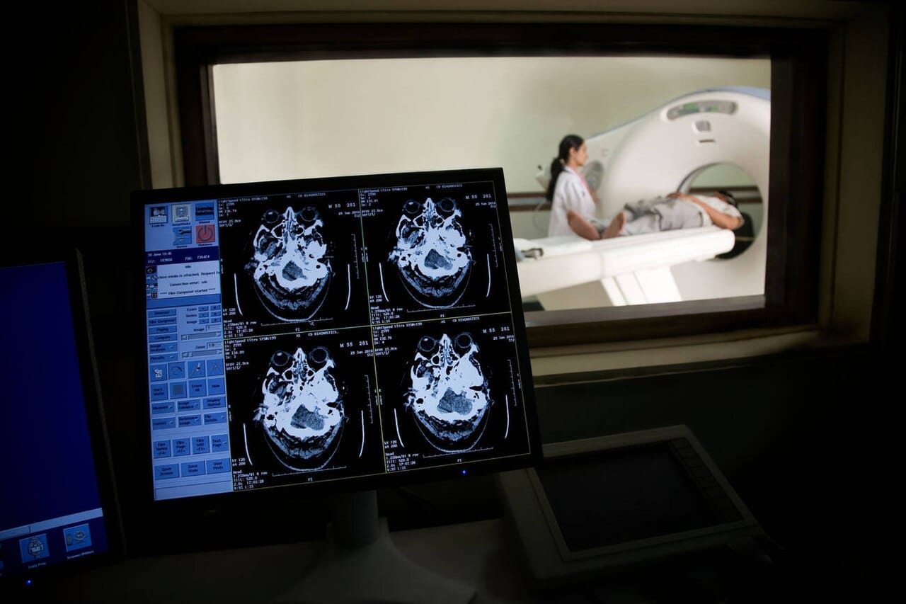
Computed tomography takes X-ray measurements from many angles and reconstructs cross-sectional images. It is fast, widely available and good at detecting bleeding and fractures, which makes it the standard first investigation in emergencies. It uses ionising radiation and resolves soft tissue less well than the alternative.
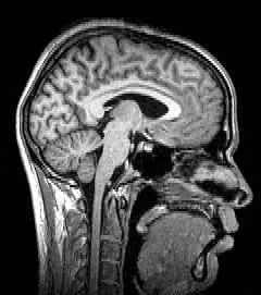
Magnetic resonance imaging places the head in a strong magnetic field and uses radio pulses to detect signals from hydrogen nuclei, which differ between tissue types. It gives far better soft tissue contrast than computed tomography, uses no ionising radiation, and is slower, more expensive and unsuitable for patients with certain implants.
Diffusion imaging measures the direction in which water moves within tissue, which follows nerve fibre bundles, and allows white matter tracts to be mapped.
Functional magnetic resonance imaging is the dominant research tool, and what it measures is indirect.
It detects the blood oxygen level dependent signal. Active neurons consume oxygen, local blood flow increases in response, and the increase overshoots demand, changing the magnetic properties of blood. The scanner detects that change.
This means the technique measures blood flow as a proxy for neural activity, with a delay of several seconds, and with spatial resolution of a few millimetres, which is a volume containing hundreds of thousands of neurons.
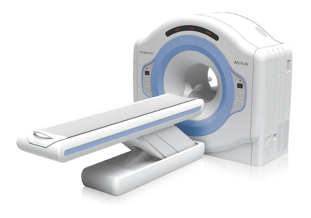
Positron emission tomography injects a radioactive tracer and detects its distribution. It can image glucose metabolism or, with appropriate tracers, the density of specific receptors or the presence of particular proteins, which is why it is central to Alzheimer's research. It involves a radiation dose and lower spatial resolution.
Electroencephalography records electrical activity from scalp electrodes, with millisecond timing and poor spatial localisation, since the signal is blurred by skull and scalp. Magnetoencephalography records the associated magnetic fields with better localisation and requires far more expensive equipment.
Functional near-infrared spectroscopy measures blood oxygenation through the skull using light, is portable and tolerant of movement, and reaches only the outer cortex.
The general trade is between temporal and spatial resolution. Electrical methods are fast and imprecise about location; blood-flow methods are precise about location and slow.
Functional images are not photographs of the brain thinking, and the gap between what is measured and what is reported is the field's persistent problem.
The pictures are statistical maps. Voxels where the signal differs between conditions beyond a threshold are coloured, and the colours are superimposed on an anatomical image. Nothing lights up; a statistical comparison exceeded a criterion.
Everything depends on the comparison chosen. An image showing a region active during a task shows activity relative to whatever the control condition was, and changing the control changes the result.
Reverse inference is the commonest error. Observing that a region activates during a task does not establish that the mental process associated with that region is occurring, because most regions participate in many processes.
The multiple comparisons problem is severe, since a brain volume contains tens of thousands of voxels and testing each produces false positives by chance. A widely cited demonstration found apparent activation in a dead salmon when the correction for multiple comparisons was omitted, which was published to make exactly this point.
Statistical practice has been substantially revised. Analyses of the software used found that some standard methods produced inflated false positive rates, and small sample sizes have been shown to yield unreliable results, with the field moving toward larger samples and pre-registration.
Clinically, structural imaging identifies tumours, strokes, bleeding, atrophy and structural abnormalities, and it changed neurology from a discipline of inference from symptoms to one with direct observation.
In research, imaging has mapped functional organisation extensively, established the existence of consistent networks including the default mode network active during rest, and allowed lesion studies to be complemented by observation of intact brains.
Presurgical mapping locates language and motor areas in individual patients before operations, which is a direct clinical application of functional methods.
Brain imaging made the living brain observable, which is a genuine transformation of both medicine and psychology.
Its interpretation is also where the public understanding of neuroscience is weakest. Brain images are unusually persuasive, and studies have found that including one increases the credibility of an accompanying claim regardless of whether it supports it, which is a reason for stating plainly what the pictures actually represent.