A form of light too energetic to see, discovered by accident in 1895, which passes through flesh and is stopped by bone. Within months it was in clinical use across the world, and it took decades to establish that it also causes cancer.
Wilhelm Rontgen was working with a Crookes tube, a partially evacuated glass tube through which an electrical discharge is passed, in November 1895. He had covered it in black cardboard to block visible light.
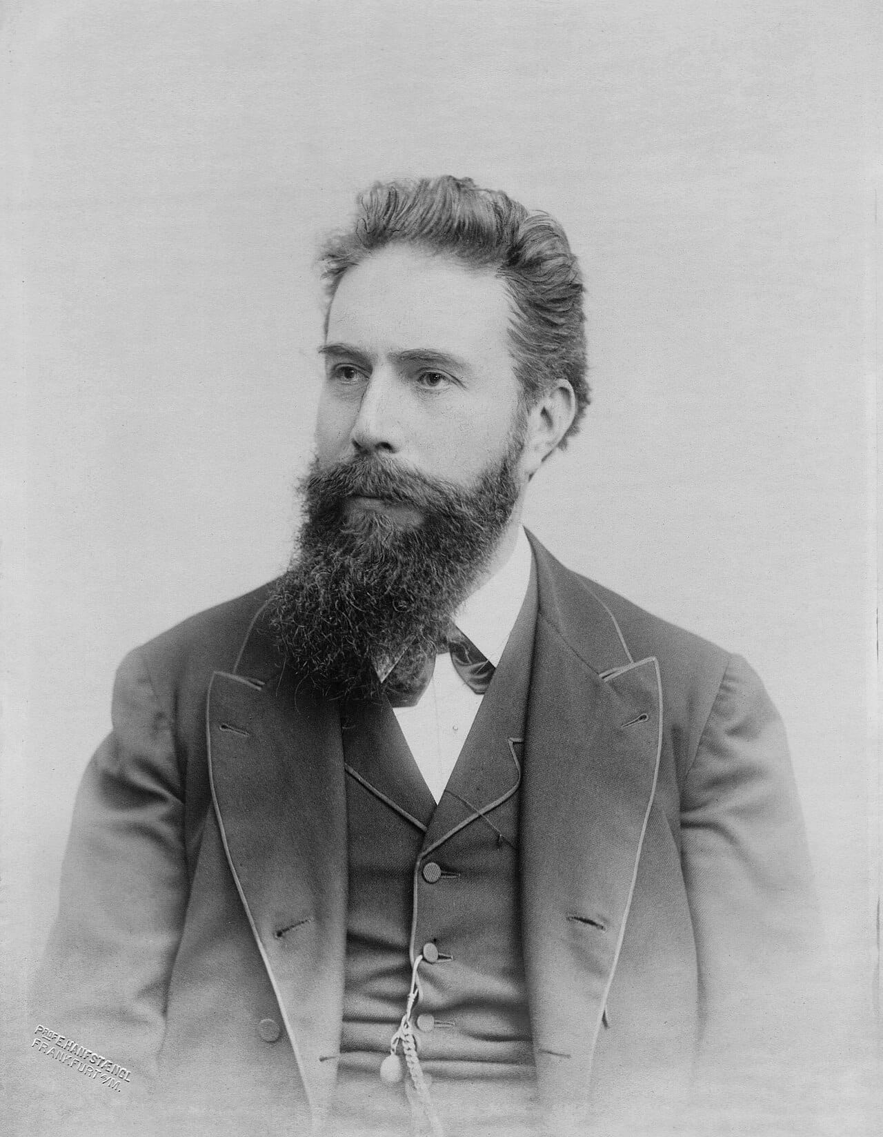
A screen coated in a fluorescent salt, sitting at some distance, glowed anyway. Something was passing through the cardboard.
He worked alone for weeks, established that the rays travelled in straight lines, were not deflected by magnets, and passed through materials in proportion to their density. He called them X-rays because he did not know what they were, and the placeholder name stuck.
He then imaged his wife's hand, producing the photograph showing her bones and her wedding ring, and published in December 1895. Her reported reaction was that she had seen her death.
The news spread with extraordinary speed. Within weeks the technique was in use in hospitals across Europe and North America, and within months it was being used to locate bullets and fractures in the field. Rontgen received the first Nobel Prize in Physics in 1901 and refused to patent the discovery, saying it belonged to humanity.
X-rays are electromagnetic radiation, the same phenomenon as visible light, radio and infrared, differing only in wavelength. Their wavelengths are around a tenth of a nanometre, comparable to the spacing between atoms, which is why they diffract from crystals and why crystallography works.
Max von Laue demonstrated the diffraction in 1912, which settled two questions at once: that X-rays are waves, and that crystals are regular lattices of atoms.
They are produced by accelerating electrons into a metal target. Two mechanisms contribute: electrons decelerating in the field of a nucleus emit a continuous spectrum, and electrons knocking inner-shell electrons out of target atoms produce sharp characteristic lines when outer electrons fall into the vacancies.
Absorption depends steeply on atomic number, roughly as its cube in the relevant energy range.
Calcium in bone has a substantially higher atomic number than the carbon, hydrogen, nitrogen and oxygen of soft tissue, so bone absorbs far more and casts a shadow. The image is a shadow, not a photograph.
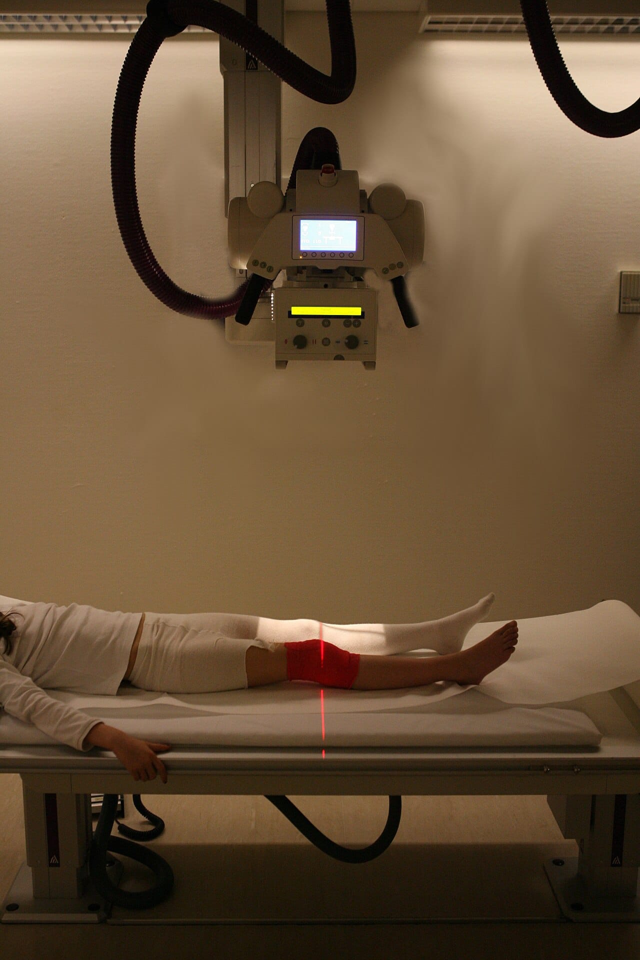
This also explains the technique's principal limitation. Soft tissues of similar density are nearly indistinguishable, which is why contrast agents containing iodine or barium are introduced to outline blood vessels or the digestive tract, and why magnetic resonance imaging is preferred where soft tissue detail matters.
Computed tomography, developed by Godfrey Hounsfield and Allan Cormack and recognised with the 1979 Nobel Prize, takes many projections from different angles and reconstructs a cross-section mathematically, which removes the superposition that makes a plain film hard to read.
The rays were treated as harmless for years and used casually. Shoe shops installed fluoroscopes for fitting children's footwear, in use into the 1970s in some places. Operators demonstrated tubes on their own hands routinely.

The consequences accumulated. Clarence Dally, Thomas Edison's assistant, developed severe burns, underwent multiple amputations and died in 1904, and Edison abandoned the work. Many early radiologists lost fingers and hands and died of cancers, and monuments to them exist in Hamburg and elsewhere.
X-rays are ionising: they carry enough energy to strip electrons from atoms, which damages DNA directly and through the free radicals produced in water. The damage is usually repaired and occasionally is not, and an unrepaired or misrepaired break can initiate a cancer.
Risk from a single diagnostic examination is small and not zero. The doses are managed on the principle of keeping exposure as low as reasonably achievable, with shielding, collimation, faster detectors and, importantly, not performing examinations that will not change management. Computed tomography delivers substantially higher doses than plain radiography, and its growth has made cumulative population exposure a real public health question.
Crystallography, which determined the structures of DNA, penicillin and countless proteins.
Astronomy, through telescopes above the atmosphere, since X-rays from space are absorbed before reaching the ground. X-ray astronomy is how black holes, neutron stars and the hot gas in galaxy clusters are studied.
Security screening, industrial inspection of welds and castings, and examination of paintings, where an X-ray frequently reveals an earlier composition beneath the visible one.
Radiotherapy, which uses the same ionising damage deliberately, delivering it to a tumour from many directions so that the target receives a lethal dose while surrounding tissue receives much less.