A way of photographing the inside of a living body in any plane, at high resolution, without ionising radiation, by making hydrogen nuclei resonate in a magnetic field. It is the most physically sophisticated instrument in routine clinical use.
Protons behave as tiny magnets. In a strong magnetic field they align with it, and they precess, wobbling about the field direction like a spinning top, at a frequency proportional to the field strength.
A radio pulse at exactly that frequency tips them out of alignment. When the pulse stops they return, emitting a radio signal as they do. This is nuclear magnetic resonance, described independently by Felix Bloch and Edward Purcell in 1946, for which they shared the 1952 Nobel Prize in Physics.
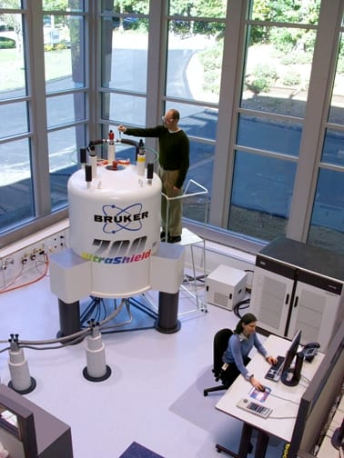
Chemists had used the effect for structural analysis for two decades before anyone imaged with it.
The obstacle was that resonance frequency depends only on field strength, so a uniform field gives one signal from the whole sample and no spatial information at all.
Paul Lauterbur's insight in 1973 was to apply a deliberate field gradient, making the field vary across the sample. Frequency then encodes position: a proton on one side resonates faster than a proton on the other. Collecting signals with gradients in several directions allows the distribution to be reconstructed, using the same mathematics as computed tomography.

Peter Mansfield developed the mathematical analysis and, critically, echo-planar imaging, which acquires a whole slice from a single excitation and reduced scan times from hours to fractions of a second. The two shared the 2003 Nobel Prize in Physiology or Medicine.
Raymond Damadian had shown in 1971 that tumours and normal tissue have different relaxation times, and held an early patent. His exclusion from the prize was publicly disputed, and he took out newspaper advertisements protesting it.
The images are of hydrogen, which is abundant in water and fat and therefore everywhere in soft tissue. What produces contrast is not how much hydrogen is present but how quickly the protons return to alignment, and that depends on the molecular environment.
Two relaxation times are measured. T1 describes recovery of alignment with the main field; T2 describes loss of coherence between protons. Different tissues have characteristically different values, and the pulse sequence can be arranged to emphasise either.
This is why MRI sees soft tissue so much better than X-ray methods, which measure only density and cannot distinguish tissues of similar density. Brain grey and white matter, ligaments, cartilage, tumour margins and inflammation are visible on MRI and largely invisible on plain radiography.
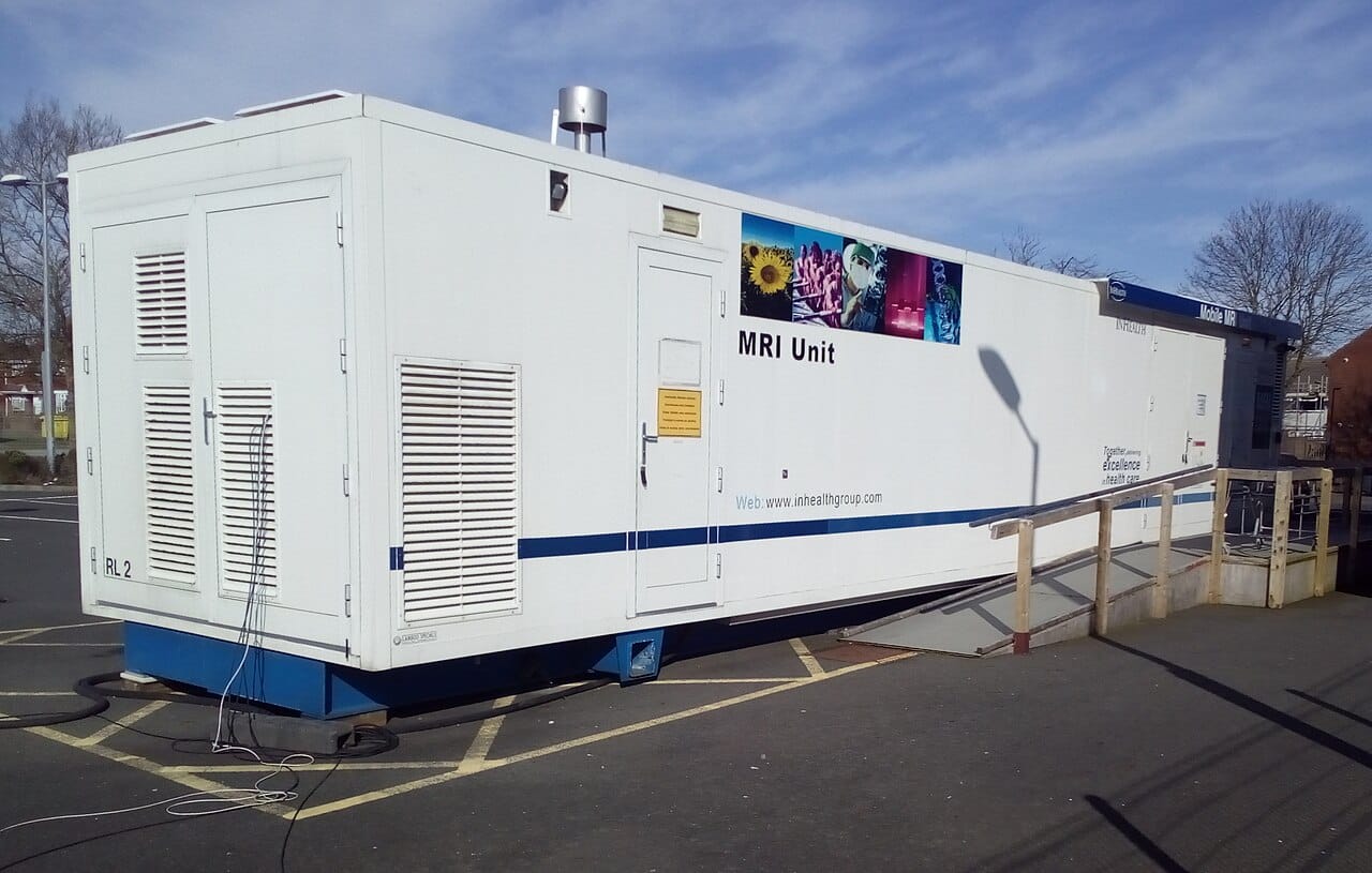
Several techniques extract information no other method provides non-invasively.
Diffusion imaging measures the movement of water molecules. Because water diffuses preferentially along nerve fibres, this maps white matter tracts through the living brain. It also detects stroke within minutes, far earlier than any structural change appears.
Functional MRI detects changes in blood oxygenation that follow neural activity, which is the basis of most human brain mapping research. The signal is indirect, measuring blood flow rather than neurons, and its interpretation requires care.
Magnetic resonance angiography images blood vessels using flow itself, frequently without any injected contrast.
Spectroscopy measures chemical composition within a chosen volume, detecting metabolites directly.
The magnet is superconducting, cooled by liquid helium, and typically 1.5 or 3 tesla, tens of thousands of times the Earth's field. It is never switched off, which is why ferromagnetic objects entering the room become projectiles and why this remains a recurring cause of serious injury.
Scans are slow compared with computed tomography, minutes rather than seconds, which makes them poor for trauma and difficult for patients who cannot lie still. The bore is confined and loud, and claustrophobia prevents a meaningful minority of patients from completing a scan.
Cardiac pacemakers and other implants were long an absolute contraindication, though many modern devices are now conditionally safe.
And the machines are expensive to buy and to run, which is the main constraint on availability and the reason access varies enormously between health systems.