A technique for determining the arrangement of atoms in a solid by firing X-rays at a crystal and reading the pattern they scatter into. It has produced more Nobel Prizes than any other method, and most of what is known about the structure of matter at atomic scale came from it.
X-rays have wavelengths comparable to the spacing between atoms, roughly a tenth of a nanometre, which is the condition for a wave to be scattered informatively by a structure. Visible light, with wavelengths thousands of times longer, passes over atomic detail without registering it, which is why no microscope using light can ever resolve an atom.
A crystal is a lattice repeating the same arrangement of atoms in three dimensions. When X-rays strike it, each atom scatters them, and the scattered waves interfere. In most directions they cancel. In specific directions determined by the lattice spacing they reinforce, producing intense spots.
William Lawrence Bragg formulated the relationship in 1912: reinforcement occurs when the extra distance travelled by waves reflecting off successive planes of atoms equals a whole number of wavelengths. Bragg's law is a single equation relating the angle of a spot to the spacing of the planes producing it, and it converts a photograph of spots into a set of distances.
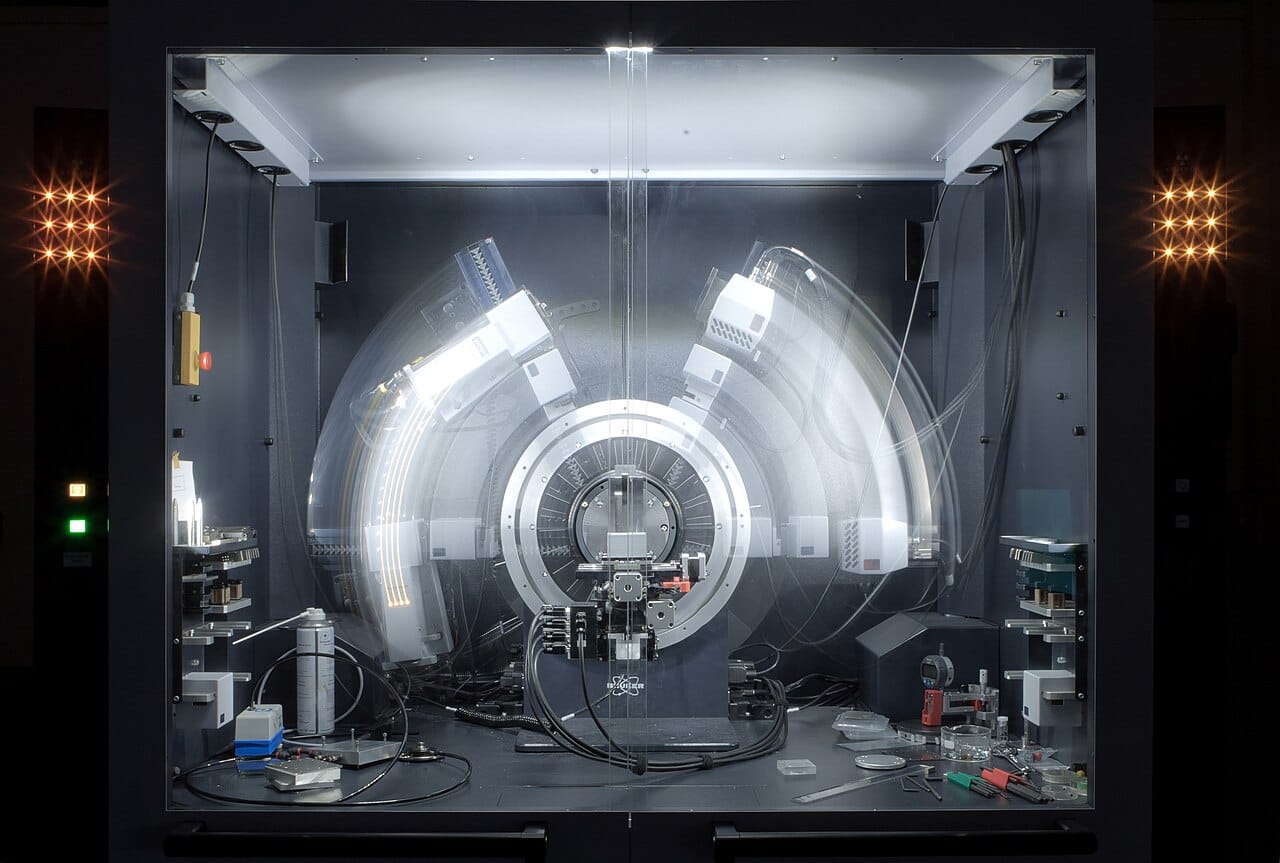
The first structures, published by the Braggs from 1913, settled arguments that chemistry could not resolve on its own. Table salt was shown to contain no discrete molecules at all: sodium and chloride ions alternate in a lattice, and the formula describes a ratio rather than a unit. Diamond was shown to be a single covalent network, which explains its hardness directly.
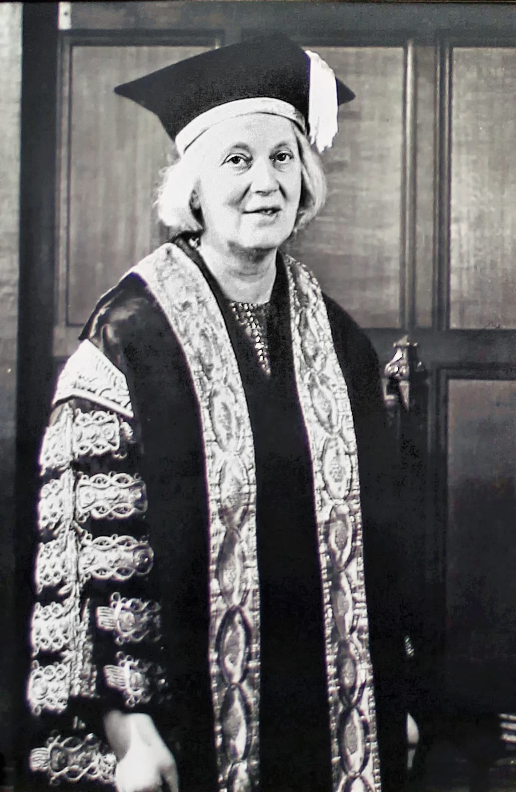
The technique then determined the structures of metals, minerals, and eventually molecules of great complexity. Dorothy Hodgkin solved penicillin in 1945, working during wartime when the structure was urgently wanted for synthesis, and vitamin B12 in 1956, a molecule with over ninety atoms whose structure had defeated conventional chemical analysis entirely. She received the 1964 Nobel Prize in Chemistry, and later solved insulin after more than thirty years of work on it.
The best known application is Photograph 51, taken in Rosalind Franklin's laboratory at King's College London in 1952 by her and the graduate student Raymond Gosling.
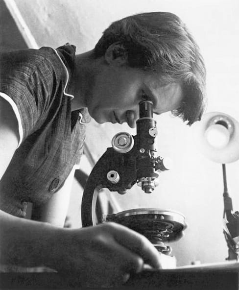
Its X-shaped pattern is the signature of a helix, and the spacing of the marks gives the pitch and diameter directly. Franklin's measurements also established that the phosphate backbone lay on the outside, which ruled out the arrangement Watson and Crick had proposed in their first, failed model.
The photograph was shown to Watson by Maurice Wilkins without Franklin's knowledge, and a report of her unpublished data reached Crick through a research council document. The 1953 structure rests substantially on her work. She died in 1958 at thirty-seven, and the Nobel Prize awarded in 1962 could not be given posthumously.
The central technical difficulty is that a detector records the intensity of each diffracted beam but not its phase, and reconstructing a structure requires both. Half the information is lost at the moment of measurement.
Solving it occupied decades. Isomorphous replacement compares patterns from crystals with heavy atoms substituted at known positions. Anomalous scattering exploits how atoms scatter differently near an absorption edge. Direct methods, developed by Jerome Karle and Herbert Hauptman and recognised with the 1985 Nobel Prize, use statistical relationships among the intensities to recover phases computationally.
The method's principal limitation is that it requires a crystal, and many important molecules resist crystallising, membrane proteins most notoriously. Growing a usable crystal has often taken longer than solving the structure from it.
Cryo-electron microscopy has taken over much of structural biology since 2013, since it works on frozen solutions and needs no crystal. AlphaFold now predicts many protein structures computationally. Neither displaces crystallography for small molecules, minerals and materials, where it remains the definitive method and where the resolution it achieves is still unmatched.