Muscle shortens because two sets of protein filaments slide past one another, driven by myosin heads that repeatedly attach to actin, pivot, and let go, each cycle consuming one molecule of ATP. The filaments themselves do not change length, which is the observation the mechanism is named for.
Skeletal muscle is built in nested repeating units, and the regularity is what made the mechanism visible.
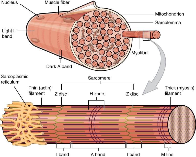
A muscle is a bundle of fascicles, each a bundle of muscle fibres, each fibre a single multinucleated cell packed with myofibrils. A myofibril is a chain of sarcomeres, the contractile unit, bounded by Z discs.
Within a sarcomere, thin filaments of actin anchor to the Z discs and thick filaments of myosin lie between them. The alternating overlap of thick and thin filaments produces the light and dark bands that give the tissue its striated appearance.
In 1954 two independent groups, Andrew Huxley with Rolf Niedergerke and Hugh Huxley with Jean Hanson, published in the same issue of Nature the observation that settled the question.
During contraction the dark band stays the same width while the light bands and the distance between Z discs shrink. If the filaments themselves shortened, both bands would narrow. They do not, so the filaments must be sliding past each other.
The cross-bridge cycle is how the sliding is driven. A myosin head carrying the products of ATP hydrolysis binds actin, releases phosphate and undergoes a conformational change that pulls the thin filament past the thick one, then releases ADP. Binding a fresh ATP detaches the head, which is then re-cocked and can attach again further along.
Two consequences of that sequence are worth stating because they are testable. Force is proportional to how many heads are attached at any instant, which is why the length tension curve peaks at the overlap that puts the most heads in reach. And ATP is required to let go rather than to hold on, which is why muscle stiffens after death: rigor mortis is myosin locked to actin with no ATP left to release it.
The link between electrical signal and mechanical output is calcium, and every step has been isolated.
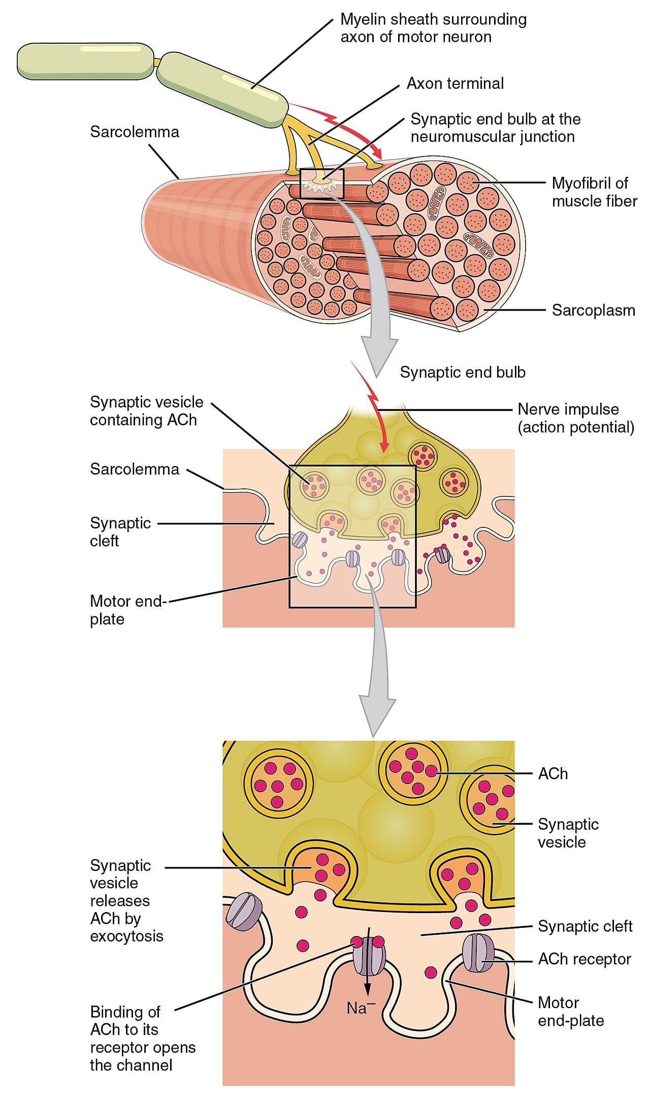
A motor neuron releases acetylcholine at the neuromuscular junction, depolarising the muscle fibre. The depolarisation travels along the membrane and inward through the transverse tubules.
Voltage sensors in the tubule membrane are mechanically coupled to calcium release channels in the sarcoplasmic reticulum, which opens them, flooding the cytoplasm with calcium.
At rest, tropomyosin lies across the myosin binding sites on actin. Calcium binds troponin, troponin shifts tropomyosin aside, and the sites become available. Contraction is therefore switched on by removing a block, not by activating the motor.
Relaxation requires pumping calcium back into the sarcoplasmic reticulum against its gradient, which costs ATP. A substantial fraction of resting muscle energy use goes on this, and it is why relaxation is an active process that can fail.
A single action potential produces a twitch. Force is graded in two ways: by recruiting more motor units, and by firing them faster so that twitches sum, up to a smooth maximal tetanus.
Motor units are recruited in a consistent order from small to large, which is why fine control is available at low forces.
Cardiac muscle uses the same sliding filament machinery and the same troponin switch, but its cells are electrically coupled and it generates its own rhythm, so it contracts as a unit and cannot be tetanised.
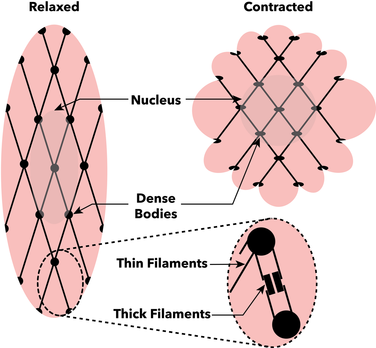
Smooth muscle, in blood vessels, gut and airways, is regulated on the myosin rather than on the actin. Calcium binds calmodulin, which activates myosin light chain kinase, which phosphorylates myosin to let it cycle. Its latch state lets it maintain tension with very little ATP, which is what a blood vessel wall needs.
The sliding filament mechanism was one of the first cases where a large-scale biological behaviour was traced to the shape changes of identified protein molecules. It also underpins clinical practice: cardiac troponin released from damaged heart muscle is the standard blood test for a heart attack, and neuromuscular blocking drugs used in anaesthesia act at the junction described above.