Proteins produced by the immune system that bind specifically to a target and mark it for removal. Their variety is generated at random before any exposure, which is how the body prepares for threats it has never met.
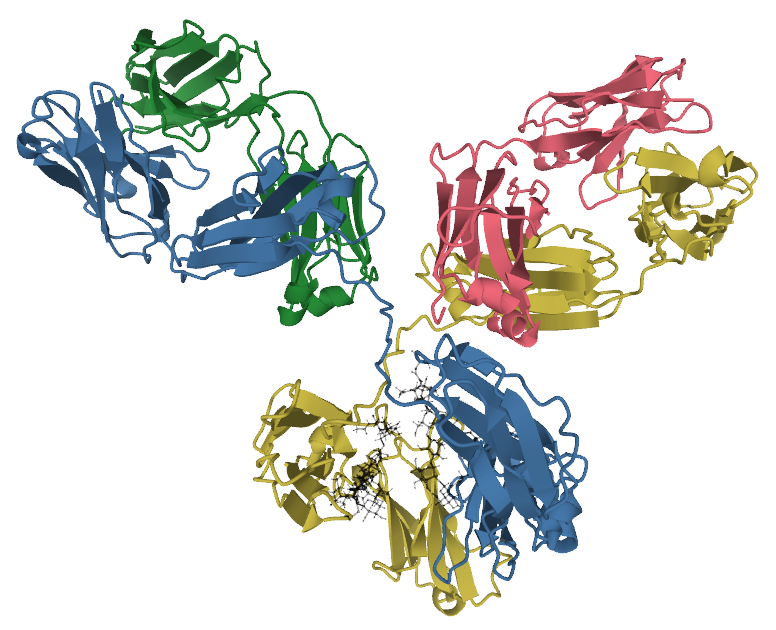
An antibody is built from four protein chains: two identical heavy chains and two identical light chains, forming a Y.
The two arm tips carry the binding sites, which are identical to each other, so one antibody binds two copies of its target. This allows it to cross-link targets into clumps, which is one of the ways it works.
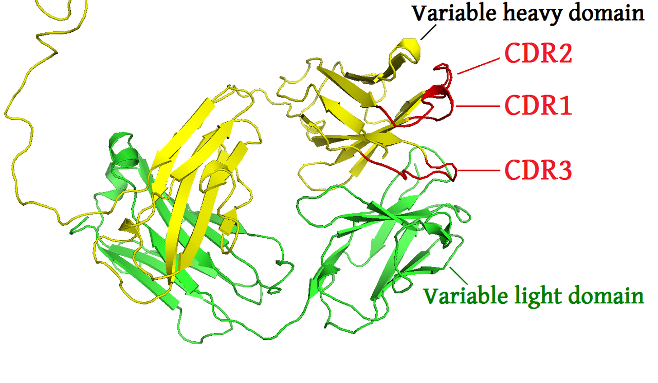
Each chain has a variable region at the tip and a constant region for the rest. Variation is concentrated in short loops within the variable region, which form the surface that contacts the target.
The constant region of the heavy chain determines the antibody's class and therefore what happens after binding, since it is the part recognised by other components of the immune system.
The specific part of a target that an antibody binds is called an epitope, and it is usually a small patch on a surface rather than a whole molecule, which is why several different antibodies can bind the same protein at different places.
The problem is severe: an organism must be able to produce antibodies against targets that did not exist when it was born, and the genome is far too small to encode a separate gene for each.
The solution is recombination. Antibody genes exist as separate segments, and each developing B cell selects one of each type and joins them, in a process that also adds and removes bases at the junctions.
The number of combinations is enormous, and each B cell ends up with one rearranged gene and therefore one specificity. Susumu Tonegawa demonstrated this mechanism and received the Nobel Prize in 1987.
Specificity is therefore generated before exposure and at random. When a target appears, the few cells that happen to bind it are selected and multiply, which is clonal selection, treated in its own capsule.
Binding then improves during the response. Dividing B cells mutate their antibody genes at a high rate, and those whose antibodies bind better are preferentially selected, a process called affinity maturation. This is variation and selection operating within one body over days.
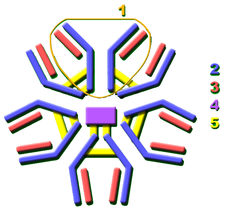
IgM is produced first in a response. It assembles into a pentamer with ten binding sites, which compensates for the relatively weak binding of individual sites early on.
IgG is the most abundant in blood and the main antibody of a mature response. It crosses the placenta, which gives newborns protection from their mother's previous exposures for several months.
IgA is secreted onto mucosal surfaces and into breast milk, defending the gut, airways and other surfaces where most pathogens first arrive.
IgE is present in small amounts, binds mast cells, and triggers the release of histamine. It defends against parasites and is responsible for allergic reactions, which is why allergy is an IgE-mediated condition.
IgD is present on B cell surfaces and its function is less well established.
Binding alone is sometimes sufficient. Neutralisation occurs when an antibody blocks the part of a virus or toxin that attaches to cells, preventing entry.
Opsonisation marks a target for phagocytes, which have receptors for the antibody's constant region and engulf what is coated.
Complement activation triggers the protein cascade that punctures bacterial membranes and recruits immune cells.
Agglutination clumps targets together through the two binding sites, which immobilises them and makes clearance easier.
Antibody-dependent cellular cytotoxicity directs natural killer cells to destroy a coated cell.
Antibodies are the most widely used reagent in biology, because they provide a way to detect one specific molecule in a complex mixture.
Diagnostic tests use them to detect pathogens, hormones and markers. Pregnancy tests and lateral flow tests are antibody-based, and blood typing uses antibodies against the relevant surface molecules.
Laboratory methods including western blotting, immunohistochemistry and flow cytometry all depend on them to identify specific proteins or cell types.
Therapeutic use is treated in the monoclonal antibodies capsule.
Detecting antibodies is also how past exposure is established, since their presence indicates the immune system has met a pathogen, which is what serological surveys measure.
Antibodies solve a problem of anticipation: preparing for an effectively unlimited set of threats using a finite genome, by generating diversity at random and selecting what turns out to be useful.
They are also the component of immunity that vaccination exploits, and the reagent on which a large part of modern biology and diagnostics depends, which makes them among the most consequential molecules both inside the body and in the laboratory.