The process that seals a damaged blood vessel, combining a physical plug of platelets with a mesh of fibrin produced by a cascade of enzymes. It has to be fast enough to stop bleeding and restrained enough not to fill the vessel, and the mechanism that achieves both is understood in detail.
Haemostasis is usually described in two parts, which happen together rather than in sequence.
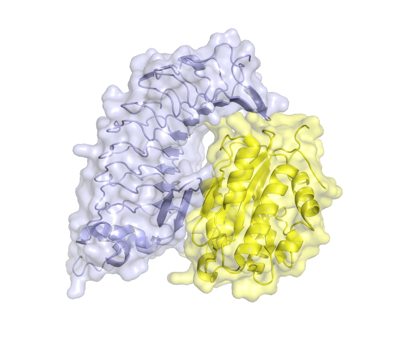
Primary haemostasis is the platelet plug. Intact vessel walls keep the underlying collagen out of contact with blood. When the lining is breached, collagen and von Willebrand factor are exposed, platelets stick to them, change shape, release granules that recruit more platelets, and aggregate into a plug.
Secondary haemostasis is the fibrin mesh. A series of circulating enzyme precursors activate one another in order, ending with the conversion of soluble fibrinogen into insoluble fibrin, which is then cross-linked into a stable network holding the platelet plug together.
A platelet plug alone is fragile and a fibrin mesh alone has nothing to build on, which is why disorders of either arm produce bleeding.
The clotting factors are mostly serine proteases circulating as inactive zymogens, each activating the next.
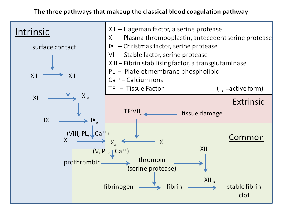
The arrangement is an amplifier. One activated enzyme cleaves many molecules of the next, so a small initiating signal becomes a large amount of thrombin within seconds. Several steps require calcium and a phospholipid surface, which the activated platelets provide, so the amplification is confined to the site of injury rather than spreading through the circulation.
Thrombin is the central enzyme. It converts fibrinogen to fibrin, activates factor XIII to cross-link that fibrin, activates platelets, and activates factors V, VIII and XI, feeding back to accelerate its own production.
Counter-regulation is equally important. Antithrombin inactivates thrombin and factor Xa, protein C and protein S shut down factors V and VIII, and tissue factor pathway inhibitor blocks the initiating complex. Clotting is a balance between two opposed sets of enzymes, not a switch.
The intrinsic and extrinsic pathways of the textbook diagram were inferred from clotting tests in glass tubes, and they describe those tests well.
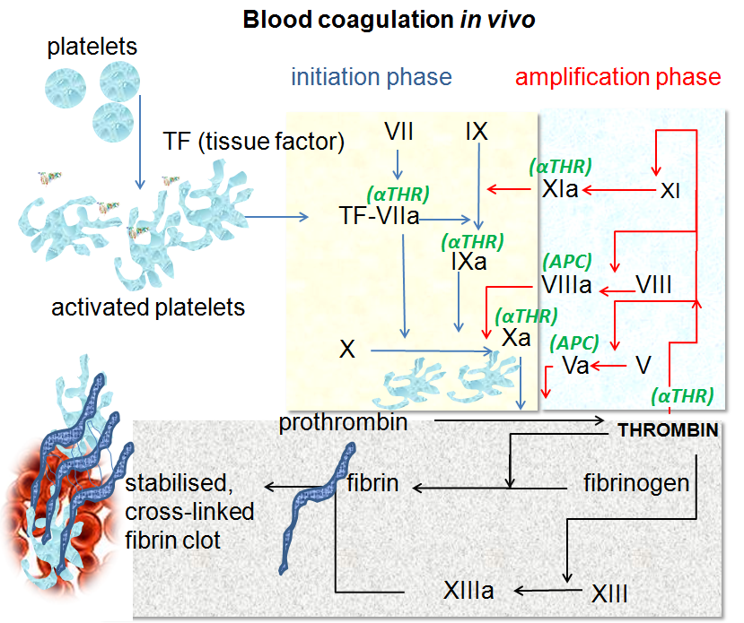
In a living vessel, clotting is initiated when tissue factor, normally absent from the blood, is exposed at the site of injury and binds factor VIIa. That complex generates a small amount of thrombin, which then amplifies the response through factors XI, VIII and V.
The clearest evidence that the contact pathway is not the physiological trigger is clinical. People deficient in factor XII have a markedly abnormal laboratory clotting time and do not bleed abnormally. Prekallikrein and high molecular weight kininogen deficiencies behave the same way.
The classical diagram survives because it maps onto the two standard tests: the prothrombin time interrogates the tissue factor arm, the activated partial thromboplastin time the contact arm. It is a good guide to interpreting a laboratory result and a poor picture of a wound.
Anticoagulant drugs are among the clearest demonstrations that the mechanism is correctly understood, because each acts at a known point and produces the predicted effect.
Warfarin blocks the vitamin K dependent modification that factors II, VII, IX and X require to bind calcium and phospholipid. It acts on synthesis, so it takes days to work and days to wear off.
Heparin accelerates antithrombin by several orders of magnitude, acting on enzymes already present, so it works within minutes.
The direct oral anticoagulants bind a single target: rivaroxaban, apixaban and edoxaban inhibit factor Xa, dabigatran inhibits thrombin itself.
Antiplatelet drugs attack the other arm. Aspirin irreversibly acetylates cyclooxygenase in platelets, which cannot make more of it, so a single dose affects a platelet for its whole lifespan.
Haemophilia A is deficiency of factor VIII and haemophilia B of factor IX, both X linked, and both produce bleeding into joints and muscles rather than from small cuts, because the platelet plug forms and then fails.
Von Willebrand disease is the commonest inherited bleeding disorder and affects the platelet arm, so it presents with mucosal bleeding and bruising.
The opposite failure is thrombosis, clotting where nothing is damaged, which causes most heart attacks and strokes and is the reason anticoagulants are among the most prescribed drugs in the world.
Disseminated intravascular coagulation is the system running out of control throughout the circulation, consuming its own factors, so that widespread clotting and severe bleeding occur at the same time.
Clotting is the standard example of a biological cascade, and it demonstrates how proportion and locality are engineered into a chemical system: amplification for speed, surface dependence for confinement, and opposed inhibitors for control. Almost every element of the mechanism has been confirmed by a drug that blocks it or a deficiency that removes it.